The Virus Microenvironment: How Cells React When Their Neighbors Get Sick

Review by Abigail Stanton (MOL, G4)

From the perspective of a cell, every viral infection is the start of a zombie apocalypse. One day you and your neighbor are working together in your quaint body community and then the next… they’ve changed. Your unrecognizable fellow cell becomes single-minded with its new obsession: making viruses. When it has spent all of its energy, it spreads the virus to all of its neighbors, turning them, too, into virus-building zombies. 

Continue reading "The Virus Microenvironment: How Cells React When Their Neighbors Get Sick"

Bacteria in the human microbiome can inactivate the antidiabetic drug acarbose

Review written by Abigail Stanton (MOL, G2)

Even in the microscopic world, survival of the fittest can make for relentless, and creative, competition. With a limited amount of resources to go around, some bacteria will play dirty to make sure they get their fair share. Actinoplanes sp. SE50/110, a bacterium that lives in the soil, has developed a strategy to fight off competitors: by producing a specialized sugar called acarbose, it can block proteins responsible for sugar uptake and metabolism in its microbial neighbors. This inhibits the growth of other bacteria, leaving more food for Actinoplanes to enjoy. 

Continue reading

Topology helps slime bacteria form fruiting bodies

Review written by Qiwei Yu (G1, QCB)

Nature never ceases to amaze us, particularly when it comes to how biological organisms develop sophisticated and diverse strategies to survive dynamic and oftentimes hostile environments. Myxobacteria (also known as “slime bacteria”), for example, have evolved a specialized life cycle to cope with the possibility of unreliable nutrient supply. While nutrient abundance enables the bacteria to grow and proliferate, nutrient scarcity can conversely trigger a transition to a more dormant state. In response to nutrient depletion, myxobacteria cells can aggregate into fruiting bodies, three-dimensional multicellular structures of diverse colors and shapes. A subset of the cells within the fruiting bodies develop into rounded myxospores with thick cell walls, waiting for a more favorable environment to resume growth. It is truly amazing how the behavior of a large number of cells can be coordinated to achieve the rapid and dynamic process of fruiting body formation.

Continue reading

Controlling microbial growth with a light switch

Review written by Olivia Duddy (MOL, G5)

Microbes are powerful tools in the biotechnology industry. Like microscopic factories, microbes are employed to manufacture a diversity of chemical compounds, such as industrial chemicals, food products, drugs, and other biotechnology molecules, on a large scale. Given the ease of genetic engineering in microbes like Escherichia coli and Saccharomyces cerevisiae, scientists and metabolic engineers alike tinker with their metabolic capacities, or even completely rewire them, to yield high concentrations of a specific product [1]. Metabolic engineers aim to maximize the efficiency of these biosynthetic processes. High efficiency, in turn, delivers biomolecules that are more readily available and at a lower cost. Metabolic engineering applications also can be more sustainable or environmentally friendly than traditional chemical synthesis approaches [1,2]. Recently, a team of researchers in the Avalos lab, led by former Ph.D. candidate Makoto Lalwani (now postdoctoral researcher at the Wyss Institute), added an additional layer of genetic engineering to this process: They are using light as a strategy to advance biomolecule production [3].

Continue reading

Exploring the role of ADP-Ribosylation in DNA repair

Review written by Jessi Hennacy (MOL, G5)

Preserving the integrity of DNA is crucial to maintaining a cell’s functions and life cycle. However, DNA is regularly under attack by chemical and physical agents, such as toxins and UV rays from the sun, that can cause breaks in the chemical backbone that holds a strand of DNA together. This DNA damage can lead to dire consequences if left unaddressed, with effects ranging from cell death to uncontrolled cellular proliferation, which leads to cancer. Thankfully, our cells have evolved mechanisms of repairing broken DNA in order to alleviate the risks of accumulating DNA damage.  

Continue reading

Understanding the entirety of the Vibrio cholerae biofilm lifecycle: First insights into biofilm dispersal

Review written by Kimberly Sabsay (QCB, G2)

Individual cells go through life cycles, in which they grow and prepare for cell division. Similarly, in certain bacteria, groups of cells go through a synergistic cycle involving the formation and disassembly of biofilms. Biofilms are essentially groups of cells that surround themselves in layers of self-made extracellular matrix proteins and polysaccharides. A biofilm is a way for cells to protect themselves from dislocation or death by predation and are present in both beneficial and pathogenic bacterial microbiomes. The sticky extracellular matrix is a structure that pathogens such as Vibrio cholerae (the bacteria that causes Cholera) can hide in to avoid detection by immune cells and thus increase their virulence. In this way, biofilms essentially provide protective armor for bacterial cells that inhibits our ability to fight pathogenic infections. 

Continue reading

dSPRINT: Machine learning for uncovering protein-ligand interaction sites

Review written by Sara Geraghty (QCB, G3)

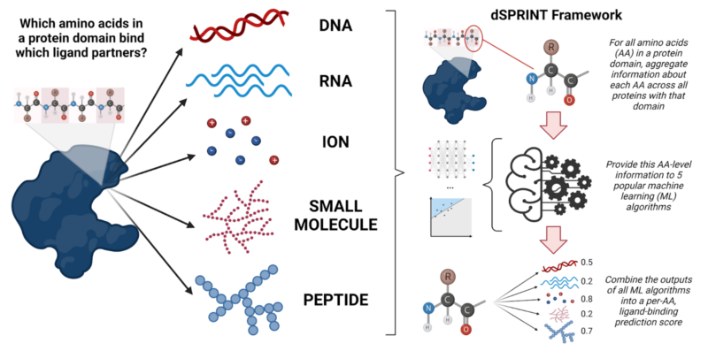
Proteins are ubiquitous in our cells. Currently, it’s estimated that the human body contains between 80,000 and 400,000 protein molecules, all busily performing the many tasks that keep your cells running smoothly and ultimately go into making you. Not only do proteins form the structural framework of your cells, but they also protect your body against foreign pathogens, help digest your food, and send and transmit signals around your body. However, they don’t do this in isolation: proteins are constantly working hand-in-hand with other proteins and molecules in your body, like your DNA, RNA, small molecules, and ions. When your DNA is replicated, or an ion is actively transported, the proteins doing the job need to recognize those molecules (and often bind to them) in order to carry out their task properly. This fundamental process of a protein binding to a partner molecule, or ligand, is critically important in biological processes ranging from development to cancer. And yet, we know surprisingly little about what proteins bind what ligands, and where -- knowledge that is key in understanding, and possibly manipulating, the inner workings of cells.

Continue reading

Structural insights into the liquid-like center of the eukaryotic CO2 concentrating organelle, the pyrenoid

Review written by Jessi Hennacy (MOL, G4)

All plants use the enzyme Rubisco to capture CO2 during photosynthesis, but Rubisco is hindered by a slow reaction rate and a counter-productive reaction that happens when the enzyme binds to oxygen instead of CO2. Algae, however, have a special organelle called pyrenoid that helps Rubisco capture CO2 more efficiently. Whereas most plants need to express high amounts of Rubisco to capture enough CO2 to grow, the pyrenoid supplies Rubisco with concentrated amounts of CO2 to improve the enzyme’s CO2 capturing activity. If a pyrenoid could be genetically engineered into crops, it could be possible for the plants to capture the same amount of CO2  with less Rubisco, thereby helping them grow with fewer resources. However, this advancement requires understanding the functional roles of proteins involved in building a pyrenoid. 

Continue reading

New tricks to study the cell's trickiest proteins

Review by Abigail Stanton (MOL, G1)

The cell can be a chaotic place to work. Protein employees of all different types rush from room to room, delivering messages, building needed materials, and working together to keep the cell running smoothly. To learn how any one of these proteins does its job, researchers have to consider how they will structure their experiment to get the type of information that they need. One approach is observing the protein at work: what does it do on a normal day? How does it interact with its coworkers? Studying a protein in situ (in its original place) gives researchers the best sense of how the protein actually behaves. However, the complex environment of the cell can make it difficult to pick out the contributions of any one protein. To gain more detailed information, the researcher may need to sit the protein down for a one-on-one interview, purifying it away from the other components of the cell for in vitro (in a test tube) experiments. However, a protein’s behavior alone may be very different from how it acts surrounded by a crowd of molecules. To create the most useful experiment possible, researchers need to find ways to combine the context of in situ studies with the detail and experimental control of in vitro work. 

Continue reading

Unlocking the Key Mechanisms of Hepatitis B Infection

Cecilia Panfil (CHM, 2022) and Alexandra Libby (PNI, GS)

Worldwide, approximately 250 million people have tested positive for the Hepatitis B virus (HBV). The virus infects the liver, causing severe damage when left untreated, such as chronic infection, liver fibrosis, liver cancer and cirrhosis. The likelihood of an adverse outcome or chronic illness is higher if the disease is contracted in childhood. Transmission can occur either through birth (i.e., the mother was infected) or close contact (e.g., sexual incourse or needle sharing for injectable drugs)1. HBV is a significant global health problem; overall, it is estimated that 650,000 people die each year from HBV related illnesses2.    

Continue reading